Does size really matter? – Scientists from 3P-Medicine Laboratory applying 10x Genomics Visium Spatial Gene Expression for human brain study
21.04.2023
Elyas Mohammadi, PhD student and Katarzyna Chojnowska, postdoc from 3P-Medicine Laboratory, supervised by Dr. Jakub Mieczkowski and Prof. Arkadiusz Piotrowski has published a paper entitled Size matters: the impact of nucleus size on results from spatial transcriptomics in the journal BMC Translational Medicine (5-year IF = 7.552, 2-year IF = 8.440). Co-authors of the publications are Anna Kostecka, Magdalena Koczkowska, Marcin Jąkalski, Natalia Filipowicz, Paweł Olszewski, Jan P. Dumański from 3P-Medicine Laboratory; Michał Bieńkowski from the Department of Pathomorphology, Justyna M. Wierzbicka and Prof. Michał A. Żmijewski from the Department of Histology, Medical University of Gdańsk. The project was conducted with the collaboration of Uppsala University (Sweden), University Health Network (Canada), University of Toronto (Canada), and Harvard Medical School (USA).
Visium Spatial Gene Expression (ST) is a cutting edge method that combines histological spatial information with direct trascriptomics profile from the tissue section. It allows to study new modes of gene expression regulations. However, it is important to remember while running ST experiment, that nucleus size may exceed the thickness of the tissue slice. As a consequence it can negatively affect comprehensive capturing the transcriptomics profile in a single slice, especially for tissues having large differences in the size of nuclei. In our work we defined the effect of Consecutive Slices Data Integration (CSDI) on unveiling accurate spot clustering and deconvolution of spatial transcriptomic spots in human postmortem brains. By considering the histological information as reference, we assessed the improvement of unsupervised clustering and single nuclei RNA-seq and ST data integration before and after CSDI. We proved that CSDI can be applied to investigate consecutive sections studied with ST in the human cerebral cortex, avoiding misinterpretation of spot clustering and annotation, increasing accuracy of cell recognition as well as improvement in uncovering the layers of grey matter in the human brain.
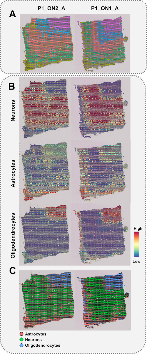
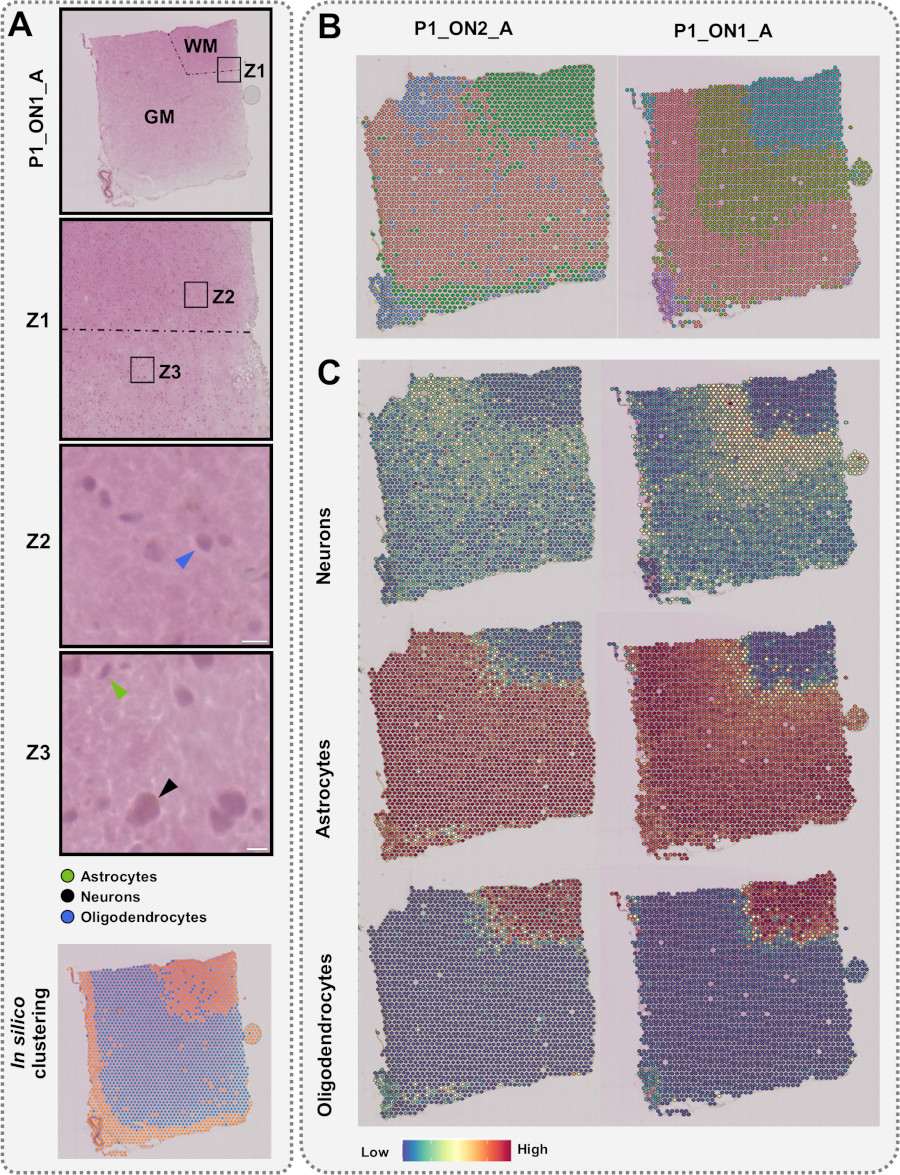


photo 3P-Medicine Laboratory/MUG & Paweł Sudara/MUG